Tooth Extraction Blood Clot Stages: What Your Socket Should Look Like
Written & Researched By
Editorial Team · product research desk
Sources: ADA guidelines, Mayo Clinic, PubMed, peer-reviewed dental literature
Published: May 25, 2026
Short answer: The blood clot in your extraction socket changes appearance as it heals. It starts as a dark red jelly-like mass on day 0, transitions to a lighter pinkish-white (granulation tissue) around days 3-5, gets covered by gum tissue between days 7-14, and the socket closes completely within 3-4 weeks. Each stage looks different, and knowing what to expect can prevent unnecessary worry.
One of the most common concerns after a tooth extraction is looking at the socket and not knowing whether what you see is normal. The color changes, the texture changes, and sometimes what’s happening in there looks alarming when it’s actually healthy healing. Here’s a visual guide to each stage.
This page walks the healing timeline twice: once as the biology your body is actually running under the surface, and once as the view in your bathroom mirror — including the white tissue that alarms almost everyone and is often a healthy sign. For photo-by-photo reference, see the tooth extraction healing stages visual guide, and for the bigger recovery picture, the tooth extraction recovery day-by-day timeline.
The Biology in Brief: Four Phases Behind What You See
Dentists and wound-care clinicians describe extraction-socket healing as four overlapping phases. You never see the phases directly — you see their side effects. Mapping them to the mirror makes every stage below predictable instead of scary.
- Hemostasis (minutes to hours). Injured blood vessels constrict, platelets pile into a plug, and the clotting cascade spins a mesh of fibrin — a stringy protein that traps red blood cells. That dark red “jelly” is mostly red cells caught in a fibrin net.
- Inflammation (days 1-3). White blood cells migrate into the clot to clear debris and bacteria. This cleanup crew is why swelling, tenderness, and pain peak on days 1-2 before turning a corner.
- Proliferation (days 3-14). The clot is gradually dissolved from within by enzymes (chiefly plasmin) while fibroblasts and sprouting capillaries build granulation tissue in its place, and epithelial cells migrate in from the gum edges to re-surface the wound.
- Remodeling (weeks to months). Osteoblasts fill the socket with hastily woven bone, which is slowly replaced by mature, organized lamellar bone over the following months.
Two insights from this biology frame everything else on this page:
- A socket never grows a skin-style scab. A scraped knee closes from the surface downward and caps itself with a hard crust. An extraction socket heals the opposite way — from the bottom and sides upward, with soft tissue doing the covering. That inside-out healing is why the socket stays moist and changes color instead of scabbing over.
- The clot is dissolved, not dropped. After roughly day 3, the clot doesn’t “fall out” — it is enzymatically dismantled as new tissue replaces it. A healthy socket should never look suddenly empty. Sudden emptiness plus surging pain is the signature of a problem, not a milestone.
Stage 1: Blood Clot Formation (Day 0, First Few Hours)
What’s happening
Immediately after the tooth is removed, blood fills the empty socket. Within 15-30 minutes of sustained pressure (biting on gauze), platelets in the blood begin aggregating and forming a clot. The clotting cascade then lays down fibrin, and within hours the socket holds a stable, structured plug — not just pooled blood.
What it looks like
- Color: Dark red, like a blob of dark jelly
- Texture: Soft and slightly raised, sitting in or slightly above the socket opening
- Size: Fills the entire socket opening
- You may see blood-tinged saliva for the first 4-6 hours — this is normal oozing, not active bleeding (bleeding after tooth extraction covers where that line sits)
What it’s doing
The clot serves as a protective cover over the exposed bone and nerve endings at the bottom of the socket. Without this clot, those structures are exposed to air, food, and bacteria, which can cause the intense pain associated with dry socket.
The clot also provides a framework. New tissue will grow through and on top of this clot structure. Think of it as the scaffolding that the rest of the healing process builds on — and, like scaffolding, it comes down piece by piece only after the building behind it is standing.
How to protect it
This stage is the most vulnerable. The clot is fresh and can be dislodged easily.
- Bite on gauze for 30-45 minutes with firm, constant pressure
- Do not rinse, spit, or use straws for 24 hours — suction is the single biggest threat to a new clot (see when it’s safe to use a straw again)
- Do not smoke — the suction and chemicals both threaten clot stability (smoking after tooth extraction explains the full risk)
- Eat only cool, soft foods on the opposite side of your mouth
- Sleep with your head elevated on a wedge pillow to reduce blood pressure at the site
Stage 2: Clot Maturation (Days 1-2)
What’s happening
The fresh clot stabilizes and begins organizing. White blood cells infiltrate the clot to clean up debris and fight bacteria. Your body’s inflammatory response peaks during this window, which is why swelling and pain are at their worst — this is the inflammatory phase of healing doing its job, not a sign that anything is wrong.
What it looks like
- Color: Still dark red, but may start developing lighter edges. Some patients notice a whitish or yellowish film on the surface — this is fibrin, the same protein mesh that holds the clot together, and it is often a normal part of healing
- Texture: Firmer than day 0, slightly less jelly-like
- The surrounding gum: Red, swollen, tender to touch (swelling peaks around now and then fades — how long swelling lasts after tooth extraction has the full curve)
Common concern: “Is that white stuff on my clot normal?”
Often, the white or yellowish material you see on the surface of the clot is fibrin. It can be part of the clot structure and a normal part of wound healing. Appearance alone cannot confirm what the material is, so if it is thick, foul-smelling, or accompanied by fever or increasing pain, call your dentist for an evaluation.
Stage 3: Granulation Tissue Formation (Days 3-5)
What’s happening
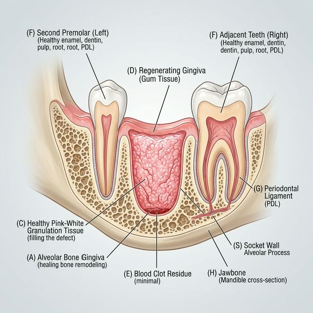
This is the most dramatic visual change. The blood clot is being gradually replaced by granulation tissue — a new type of tissue that your body builds specifically for wound repair. Fibroblasts (tissue-building cells) move in, new blood vessels sprout into the area (a process called angiogenesis), and the old fibrin mesh is dissolved from within. The scaffolding comes down as the rebuild goes up.
What it looks like
- Color: Shifting from dark red to pinkish, pinkish-white, or creamy white. The socket may look lighter than you expect.
- Texture: Soft, slightly bumpy or pebbly. Some patients describe it as looking like a wet, pink marshmallow. It’s fragile — don’t poke or prod it.
- Size: The socket is starting to shrink as the tissue fills in from the edges and bottom
For a detailed look at this phase, read granulation tissue after tooth extraction: what it is and why it matters.
Common concern: “My socket is turning white. Is this dry socket?”
This may not be dry socket. Dry socket can involve specific accompanying symptoms: severe, worsening pain that radiates to the ear or temple, a bad taste, and OTC pain meds that don’t help. If the socket is turning whitish-pink but your pain is improving daily, what you’re seeing may be healthy granulation tissue. If you are unsure or your pain is worsening, ask your dentist to evaluate it. For a full comparison, see dry socket symptoms vs normal healing.
Stage 4: Tissue Coverage (Days 5-10)
What’s happening
The gum tissue at the edges of the socket is migrating inward, gradually covering the granulation tissue beneath. The surface of the socket transitions from an open wound to a closed surface. Deeper in the socket, early bone formation is beginning.
What it looks like
- Color: Pinkish-red gum tissue growing in from the edges, with lighter tissue visible in the center
- Texture: Smoother than the granulation tissue stage. The bumpy, pebbly appearance gives way to more uniform tissue
- Size: The opening is visibly smaller than it was a week ago
- A shallow depression or indentation is normal — the socket doesn’t fill all the way to the surface in this stage
What to watch for
This is typically a low-risk stage. By now, the chances of developing dry socket are very low (dry socket almost always begins between days 2-4). Continue being gentle when brushing near the area (when you can brush normally again) and rinsing after meals with an alcohol-free rinse like CloSYS or warm salt water — more options in the best oral rinses for post-extraction healing.
Stage 5: Surface Closure (Days 10-21)
What’s happening
The gum tissue fully covers the extraction site. Beneath the closed surface, the socket continues filling with new bone tissue. The soft tissue on top is still maturing and strengthening.
What it looks like
- Color: Pink, matching the surrounding gum tissue. May be slightly more red or shiny than the adjacent gums for a few more weeks
- Texture: Smooth, continuous gum surface. The site may look slightly indented compared to the surrounding tissue
- No open wound is visible
What you can do
- Resume normal brushing and flossing in the area
- Eat normally — hard and crunchy foods are fine now for most patients
- The indentation will gradually fill in over the coming months as bone remodels underneath
Stage 6: Bone Remodeling (Months 1-6)
What’s happening
This is the longest stage but the least noticeable. Woven bone (hastily built structural bone) is gradually replaced by lamellar bone (mature, organized bone). The socket fills in completely from the bottom up.
What it looks like
From the outside, nothing. The gum surface looks normal. You may feel a slight depression with your tongue for the first month or two, but it gradually levels out. By 4-6 months, an X-ray would show fully remodeled bone filling the socket.
This stage matters most for patients planning dental implants, since an implant requires sufficient bone volume. Your dentist may have placed a bone graft at the time of extraction to preserve the socket shape for a future implant.
The White-Tissue Twin: Which White Is Worrisome?
Here is the question this whole timeline really hinges on: something white is in your socket — is it healing or trouble? At least four different things can look white in a healing socket; some may reflect normal healing, while others warrant dental evaluation. Telling the twins apart comes down to timing, texture, and above all your pain trend. (The deepest single-page treatment of this question is white tissue in the tooth extraction site; here is the clot-stage-specific version.)
Fibrin film (days 1-2)
A thin, whitish-yellow, filmy layer sitting on the clot surface. Pain is stable or improving. This often reflects clot structure doing its job, but ask your dentist to evaluate it if you have worsening symptoms.
Granulation tissue (days 3-10)
Creamy pink-white, moist, pebbly, filling the socket from the bottom and edges. Pain improves day over day. This may reflect the rebuild in progress, but ask your dentist to evaluate it if the appearance or symptoms concern you.
Food debris and slough (any day)
Soft white material — rice, bread, dairy, or fibrin slough — clinging to the site. Pain unchanged. It may gently rinse away with warm salt water after the first 24 hours. This is often harmless, but don’t pick at it.
Material that may be pus (possible infection)
Thicker, yellowish-green material, especially with a bad taste or odor, rising pain, fever, or swollen lymph nodes, may indicate infection. Call your dentist to evaluate these findings; see infection signs after tooth extraction.
Exposed bone (possible dry socket)
The socket may look empty or hollow, with whitish-gray bone visible at the base instead of tissue filling it, and severe pain that spikes on days 2-4 and radiates to the ear or temple. These findings may indicate dry socket and warrant a call to your dentist. Exposed bone can look white too, but the combination of a vacant-looking socket and pain that climbs instead of fades should be evaluated.
| White finding | Typical timing | Pain trend | Other clues | Verdict |
|---|---|---|---|---|
| Fibrin film | Days 1-2 | Stable or improving | Thin, sits on the clot surface | Often normal |
| Granulation tissue | Days 3-10 | Improving daily | Creamy pink-white, pebbly, moist | Often normal |
| Food debris | Any | Unchanged | Rinses away gently | Often harmless |
| Pus-like material | Any | Worsening | Fever, swelling, bad taste, swollen lymph nodes | Call your dentist |
| Exposed bone | Onset days 2-4 | Severe and worsening | Socket looks empty, radiating pain | May indicate dry socket — call your dentist |
Visual Quick Reference
| Stage | Days | Healing phase | Color | Texture | Pain |
|---|---|---|---|---|---|
| Blood clot formation | Day 0 | Hemostasis | Dark red | Jelly-like, soft | Moderate |
| Clot maturation | Days 1-2 | Inflammation | Dark red + white film (fibrin) | Firmer, stable | Peak |
| Granulation tissue | Days 3-5 | Proliferation | Pinkish-white, creamy | Bumpy, pebbly, soft | Improving |
| Tissue coverage | Days 5-10 | Proliferation | Pink edges closing inward | Smoother, shrinking opening | Mild |
| Surface closure | Days 10-21 | Maturation | Pink, matching gums | Smooth, continuous | Minimal to none |
| Bone remodeling | Months 1-6 | Remodeling | Normal gum color | Normal gum texture | None |
What the Stages Look Like When Something Goes Wrong
Dry socket
If the clot is missing, the socket may look hollow with whitish or grayish bone visible at the bottom instead of tissue filling it. Severe pain starting on day 2-4 that radiates to the ear or temple may indicate dry socket and warrants prompt dental evaluation — the Mayo Clinic describes pain that begins within a few days of the extraction, can travel to the ear, eye, temple, or neck, and may be accompanied by a visible empty-looking socket and bad taste or odor. The NHS likewise flags severe pain developing in the days after extraction, when the clot is lost or never forms, as the hallmark warning sign. Read the dry socket symptoms guide for the full picture, dry socket signs and prevention to lower your risk, and how to find an emergency dentist for dry socket if it lands outside office hours.
Infection
Red, angry-looking, swollen socket tissue, thick yellowish-green material, fever, increasing pain, or swollen lymph nodes may indicate infection. Ask your dentist to evaluate these signs. See infection signs after tooth extraction.
Bone spicule
A small, hard, white fragment poking through the gum tissue may be a piece of bone that broke off during extraction and is working its way to the surface. It is often uncomfortable but not dangerous; ask your dentist to evaluate it, and they can remove it in a quick visit if needed. Read more about bone spicules after tooth extraction.
Common recovery questions
What color should a healing tooth extraction look like?
Dark red on days 0-2 (blood clot), transitioning to pinkish-white on days 3-5 (granulation tissue), then gradually matching the pink of your surrounding gum tissue as the surface closes over days 7-21. The changing colors are often part of healthy progression, not necessarily a reason for concern.
Is it normal for the extraction site to be white?
Often, yes. White or pinkish-white tissue appearing on days 3-5 may be granulation tissue — a sign that your body may be building new tissue. However, if the white appearance is accompanied by severe, worsening pain and visible bone at the bottom of the socket, it may indicate dry socket and warrants a call to your dentist.
What’s the difference between fibrin and granulation tissue?
Fibrin is the protein mesh that forms and holds the blood clot — on days 1-2 it can show up as a thin whitish film on the clot’s surface. Granulation tissue is the new tissue that grows through and replaces that mesh starting around day 3: pinkish-white, pebbly, and loaded with fresh capillaries. Fibrin is the scaffolding; granulation is the rebuild. Both can be part of normal healing and both can look white.
Can food look like white tissue in the socket?
Yes. Soft white foods — rice, bread, pasta, dairy — can lodge in or against the site, and a layer of pale slough can also sit on healing tissue. If pain is otherwise improving, gently rinse with warm salt water (after the first 24 hours) rather than picking at it. Material that keeps returning, or that arrives with rising pain or a bad taste, deserves a call to your dentist.
How do I know if my blood clot fell out?
If the clot dislodges, you may taste blood or notice the socket looks empty. The most telling sign can be a sudden worsening of pain on day 2-4 that is distinctly more severe than day 1. You may see bone at the bottom of the socket instead of a dark red filling. Ask your dentist to evaluate these findings.
Can I see the blood clot in my extraction socket?
Yes, usually. Look in a mirror with a flashlight. In the first 1-3 days, you should see a dark red mass filling the hole where the tooth was. As healing progresses, the appearance changes (see the stages above), but the socket should generally look filled with tissue, not hollow.
How long does the blood clot stay in the socket?
The blood clot itself is gradually replaced by granulation tissue starting around day 3. By day 7-10, the original clot has been almost entirely replaced by new tissue. You don’t lose the clot — it transforms, dissolved from within by your body’s own enzymes as new tissue takes over. The risk of dry socket (from clot dislodgement) is highest in the first 3-4 days.
Key Takeaway
Your extraction socket goes through dramatic visual changes during healing — dark red, whitish film, pinkish-white, and finally normal pink gum tissue — and many of these changes are part of normal healing. Your pain trend can be more informative than the color of the socket. If pain is improving daily from day 3 onward, what you’re seeing may be healthy healing. If pain is worsening after day 3 — especially with an empty-looking socket, visible bone, a bad taste, or pain radiating to your ear or temple — those findings may indicate dry socket or infection rather than routine healing; call your dentist promptly so they can evaluate and treat the cause.
Disclosure: Some links on this page are affiliate links. If you make a purchase through them, we may earn a commission at no additional cost to you. This never influences what we recommend — and nothing here replaces your own dentist’s post-operative instructions.